Programa de la clase
Morfología de las gametas masculina y femenina. Transporte de gametas. Proceso de capacitación del espermatozoide: mecanismos y etapas. Concepto de reacción acrosómica. Interacciones entre gametas y reconocimiento entre espermatozoide y ovocito. Interacción del espermatozoide con las envolturas del ovocito. Penetración de la membrana pellúcida y proceso de fusión de gametas Concepto de activación del ovocito. Principales eventos durante la activación del ovocito: bloqueo de la poliespermia, reactivación de la meiosis, incremento del metabolismo. Aportes de cada gameta (masculina y femenina) al desarrollo del cigoto.
Seminarios
Gametas masculina y femenina: gametogénesis y morfología. Transporte de gametas. Proceso de capacitación del espermatozoide: mecanismos y etapas.
Objetivos mínimos
Al terminar el recorrido de la semana, los alumnos deberían ser capaces de conocer y comprender las siguientes temáticas:
- Estructura de las gametas.
- Maduración y capacitación del espermatozoide. Lugar donde ocurre y bases celulares de estos procesos.
- Ascenso y encuentro de gametas. Mecanismos que lo permiten / facilitan o impiden.
- Fusión de las membranas de las gametas. Bases celulares de la fusión y consecuencias.
- Control de la poliespermia. Bases celulares y moleculares de los mecanismos que evitan la poliespermia.
- Activación de la célula huevo e inicio del programa de desarrollo
Bibliografía destacada
Se destacan los textos mencionados sin perjuicio de que la bibliografía básica es siempre de lectura recomendable
- Capítulo 2: La fecundación, Embriología Humana de Vladimir Flores
- Capítulo 1 y 2: Gametogénesis - Transporte de los gametos y fecundación, Embriología Humana y Biología del Desarrollo de Bruce Carlson, 5ta Edición
- Capítulo 7, Biología del Desarrollo. Gilbert, 7ma Edición
Galería de imágenes
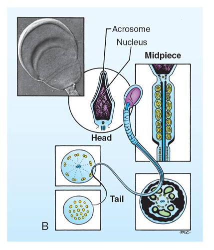
Figure 1-4, contenido. B, Structure of the mature spermatozoon. The head contains the nucleus capped by the acrosome; the midpiece contains coiled mitochondria; the tail contains propulsive microtubules. The inset micrograph shows the head of a human sperm. C, Bull sperm labeled with fluorescent markers to reveal its nucleus (blue) in its head, mitochondria (green) in its midpiece, and microtubules (red) in its tail. The red labeling around the perimeter of the head is background labeling (Larsen 4th ed)
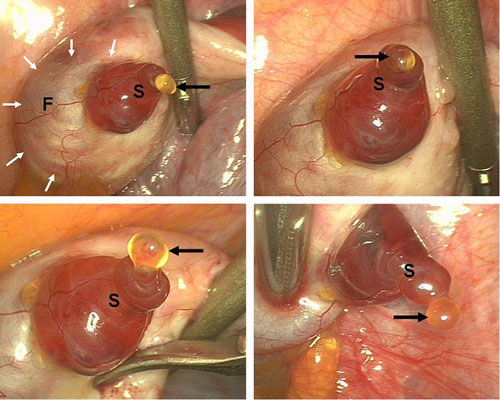
Laparoscopic observation of spontaneous human ovulation. A remarkably prominent vascular pattern was observed on the mature follicle (F, white arrows). A small follicular area called the stigma (S) was seen protruding like a reddish bleb from the follicular surface, with viscous yellow fluid (black arrows) evaginating outward into the peritoneal cavity. The viscous fluid probably carried with it the cumulus-oocyte complex, surrounded by several thousand small granulosa cells known as corona radiata.
Lousse. Laparoscopic observation of ovulation. Fertil Steril 2008. Received November 13, 2007; revised and accepted December 13, 2007. Reprint requests: Jacques Donnez, M.D., Ph.D., Department of Gynecology, Universit e Catholique de Louvain, Cliniques Universitaires St. Luc, Avenue
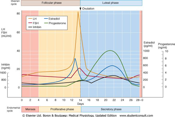
Boron, Medical Physiologhy
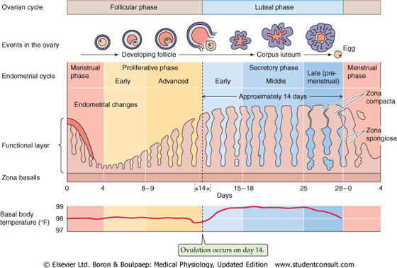
Boron, Medical Physiologhy
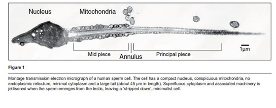
Montage transmission electron micrograph of a human sperm cell. The cell has a compact nucleus, conspicuous mitochondria, no endoplasmic reticulum, minimal cytoplasm and a large tail (about 45mm in length). Superfluous cytoplasm and associated machinery is jettisoned when the sperm emerges from the testis, leaving a stripped down minimalist cell (Barratt et al.: The human spermatozoon a stripped down but refined machine, Journal of Biology 2009, 8:63)
Gilbert, Developmental Biology
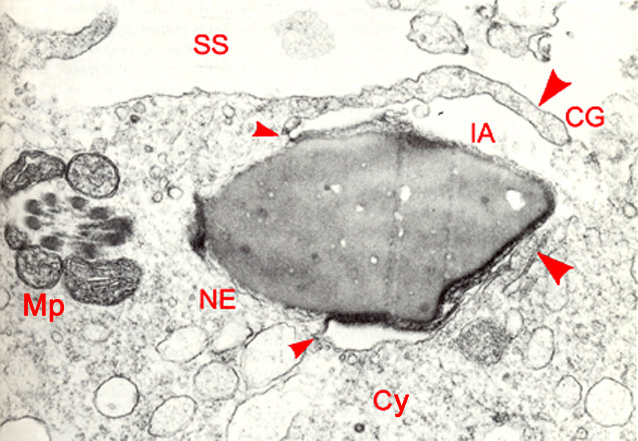
High power TEM of an early stage 1a embryo in vitro showing sperm/oocyte fusion, 2 hours post-insemination (original magnification x40,700). The sperm head has undergone the acrosome reaction and its inner acrosome membrane (IA) is exposed. The cell membrane is fused with the post-acrosomal segment of the sperm plasma membrane (small arrowheads). The front two-thirds of the sperm head is engulfed by two villous-like processes (large arrowheads) extending from the oocyte surface. The sperm nuclear envelope (NE) in the posterior region of the sperm nucleus has not yet decondensed. The plasma membrane of the sperm midpiece (Mp) has disappeared but mitochondria, dense fibers, and axoneme are still present. Cortical granules (CG) have released their products into the subzonal space (SS). The embryo is in early anaphase of the second maturation division. EHD.org
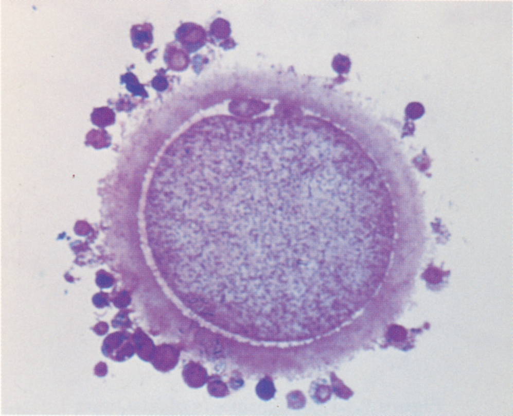
Stage 1a embryo penetrated oocyte in vitro. The second polar body is in the subzonal space indicating sperm penetration of the cell membrane. Pronuclei are absent.